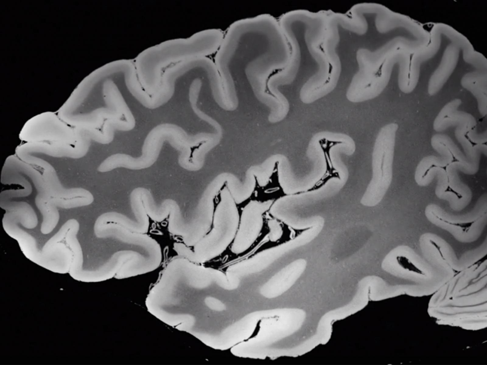
You can probably imagine our brain: a symmetrical soft mass, between pink and gray in colour, with quite a lot of bends. But until a team of researchers from the Laboratory for NeuroImaging made their recent MRI scan of a human brain (that took more than 100 hours in one of the world's most advanced MRI machines), no one had it seen in such detail — at a resolution of 100 microns, to be precise — as shown in this video:
"Thanks to an anonymous deceased patient whose brain was donated to science," writes Science Alert's Peter Dockrill, "the world now has an unprecedented view of the structures that make thought itself possible." After its extraction and "a period of preservation, the organ was transferred to a custom-built, air-tight brain holder made of rugged urethane, specially designed for the experiment's long-duration MRI scan. The holder was placed in a customized Tesla (7T) MRI scanner: a powerful machine offering high levels of magnetic field strength, and only approved by the FDA for use in the US in 2017."
Such a machine could scan a brain still in use — that is, one inside the skull of a living, breathing human being — but only for a short period of time. The great advantage of using an ex vivo brain rather than an in vivo is that it can stay inside, completely unmoving, for as long as it takes to acquire the highest-quality scan yet seen. The team could thus record "8 terabytes of raw data from four separate scan angles," data they have released to the academic community in a compressed version, which you can view and download here.
"We envision that this dataset will have a broad range of investigational, educational, and clinical applications that will advance understanding of human brain anatomy in health and disease," write the team, who are also preparing their research for publication in a peer-reviewed journal.


